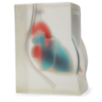
Blue Phantom’s brand-new Heart Block Model is excellent for training clinicians in the techniques associated with transthoracic echocardiography, transesophageal echo (TEE), and ultrasound guided pericardiocentesis procedural training. Blue Phantom’s TTE/TEE cardiac ultrasound training model allows users to develop and practice ultrasound imaging skills as TEE, transthoracic echocardiography, and ultrasound guided pericardiocentesis. These skills include; using ultrasound system controls, transducer insertion & placement, probe positioning and movement, applying proper transducer pressure to obtain images, recognition of the cardiac structures, guiding needles and catheters to the pericardial space for pericardiocentesis training. This portable, light weight heart model offers an extremely realistic echocardiography training platform as you utilize your own ultrasound system for simulation training. Self-healing simulated tissue allows for repeated procedural training without fluid leakage while utilizing 18 - 21gauge needles and corresponding catheters.
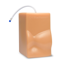
This extremely realistic, transesophageal TEE and transthoracic echo simulator is available in transparent and flesh tone configurations. Transparent version allows users to visualize the needle and the TEE probe without the use of ultrasound. This allows the user to comprehend the spatial orientation of the needle and the TEE probe. Utilizing your own ultrasound system and a TEE simulator; this simulation trainer will perform well using any ultrasound imaging system configured with appropriate transducers for echocardiography imaging procedures. Our uncompromising quality allows clinicians to utilize the model and repeatedly practice the procedure without the high costs of replacing disposable parts. Users can expect extreme durability with the simulated tissue able to perform well for thousands of pericardiocentesis procedures. This ultrasound simulation model is excellent for cardiology, anesthesiology, emergency medicine, ultrasound training programs, simulation centers, surgical skills centers, medical education facilities, and ultrasound manufacturers for ultrasound education and demonstrations.
- Heart anatomy contains left and right ventricle, left and right atria, mitral valve, tricuspid valve, aortic valve, pulmonic valve, left atrial appendage, pulmonary arteries, pulmonary veins, Pericardium, ascending aorta, SVC, and IVC for extremely realistic imaging
- Obtain images from all appropriate imaging positions including transesophageal and transthoracic imaging windows
- Excellent for 2-D, 3-D, and 4-D training
- Train clinicians to develop and practice cardiac ultrasound TEE and TTE echocardiography exams and pericardiocentesis procedures
- Extremely durable; use for repeated training without the high cost of replacing disposable parts
- Extracted fluid is easily replaced using a quick fill port or continuous filling using a luer lock
- Uncompromising image quality allows you to teach using sonographically accurate models
- Contains anatomical landmarks for the most realistic simulation training
- Practice using ultrasound system controls
- High quality, proven technology
- Patented technology
- Dimensions: 8 X 6 X 11 inches (H X W X L)
- Weight: 12 pounds